Greater Insights Faster*
The Voluson™ difference is based on dedication to Women’s Health – we partner with our customers to understand the requirements for the changing medical environment. Our customer partnerships keep us in touch with the latest trends in medical diagnosis and treatment to develop solutions to maximize imaging performance and efficiency.
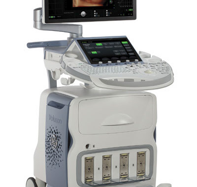
The Voluson E10 was designed with our customers in mind.

The Voluson E10 is our most advanced ultrasound system yet. With the power of the Radiance System Architecture, you can:
- Push the boundaries of extraordinary image quality for confident answers through clarity, speed and flexibility.
- Display more image detail and clarity in less time with electronic matrix 4D probe technology through ultra-fast frame rates, flexible imaging formats, and excellent resolution
- Boost exam efficiency and accuracy through Voluson automation tools
- Securely connect and share ultrasound findings with patients and colleagues
- Simplify the exam process, from patient scheduling to exam reporting, with Voluson and ViewPoint™ allowing you to focus on your patient needs
The Voluson E10 provides dedicated solutions to help address your toughest diagnostic challenges, sooner.
* Compared to Voluson Expert Series BT13
A new era of imaging performance

The Voluson E10’s Radiance System Architecture, with its sophisticated beam formation and powerful processing, sets a new standard in imaging performance to give you:
MORE CLARITY
4x Ultrasound Pathways for spectacular 2D and 3D/4D images with increased penetration.
MORE SPEED
10x the data transfer rate for higher resolution and very fast frame rates.
MORE FLEXIBILITY
4x the processing power for advanced applications and efficient workflow.
As a result, you can have extraordinary confidence in your ability to:
- Image a comprehensive range of complex women’s health issues
- Assess fetal health from the early stages
- Increase workflow efficiency and productivity
- Deliver the high level of imaging excellence your practice demands
The Radiance System Architecture of the Voluson E10 provides the robust image quality you’ve come to expect from GE Healthcare. From superb 2D images to new 3D/4D imaging technologies, you can have confidence in the accuracy of the image detail.
Simply extraordinary Electronic 4D

The Voluson E10’s Radiance System Architecture, together with the world’s first commercially available curved electronic matrix 4D probe, eM6C, delivers ultra-fast volume rates, flexible imaging formats and excellent resolution for routine OB exams to complex fetal echocardiography.
- Biplane imaging provides simultaneous display of high resolution, high frame rate images in two perpendicular planes
- VCI-A (Volume Contrast Imaging) – Delivers excellent contrast resolution through thick slice volume of grey scale and color Doppler images
- eSTIC (Spatio-temporal image correlation) – Enhances fetal cardiac exams with up to 75% reduction in acquisition time over traditional STIC
- e4D SnapShot – Optimizes your exam time with one button access from Real-time 4D to acquire high resolution 3D volume or eSTIC data sets
Your Vision – New Perspectives

More detail, more clarity in less time…. The Voluson E10 system’s new rendering technologies offers you the best image quality you have ever seen from a Voluson.
HDlive
The suite of HDlive technologies on the Voluson E10 brings unprecedented anatomical realism through advanced skin illuminating and shadowing techniques to help reveal a unique perspective for a higher level of diagnostic confidence.
- HDlive Silhouette – Dynamically apply transparency to rendered structures for a more comprehensive view of anatomy from a solid surface structure to developing internal anatomy
- HDlive Studio – Illuminate fetal anatomy with up to three independent light sources of variable intensity to focus on even the tiniest of structures
- HDlive Flow – Clearly display vascular structures with greater dimension – from small vessels to the great arteries
- HDlive Flow Silhouette – Visualize blood vessels from a surface or transparent view to provide greater insight into vascular anatomy and surrounding structures
V-SRI
Improve 3D/4D quality in multi-planar studies and enhance smoothing effect on rendered images through speckle reduction.
Advanced VCI – Adjusts slice thickness on 3D or 4D images to help enhance contrast resolution with use of render techniques such as bone and tissue renderings. Can be applied in the acquisition plane (VCI-A), static 3D volumes, or OmniView
OmniVew – Obtain any plane from a 3D or 4D volume by simply drawing a line, curve, poly-line or trace through a structure. This valuable technology enables views of even irregularly shaped structures not attainable in 2D imaging
SonoRenderlive
Enhance efficiency in volume rendering with automated placement of the render line for optimal surface rendering. SonoRenderlive continuously updates render line placement with fetal movement during 4D examinations.
Maximize efficiency with modern ergonomic design
- Cutting-edge monitor technology – high resolution. widescreen OLED monitor
- Monitor features large clipboard and standard/XL image formatting
- 12.1” Touch Panel with multi-touch functionality
- Quick and easy 1-button control panel up/down function for optimal positioning
Simplified workflow
- Electronic TGC and efficient menu navigation with swipe technology
- Bar Code scanner for efficient entry of patient information
- ViewPoint synchronization for seamless data sharing and reporting
- 4 Active probe ports with port illumination
Fast, secure data management for efficient communication
- Secure User Management – unique user IDs for system access and tracking documentation
- Integrated Software Digital Video Recorder (DVR), including USB recording
- Directly export 3D print files in multiple formats
- Archive, collaborate and share images with 3G or cloud- based connectivity
Gain Efficiency With Automation
Your day is more than just scanning. Easy-to-use automationand enhanced ultrasound workflow tools help simplify the patient exam process to address your busy practice and increase patient satisfaction.
- SonoNT – semi-automated nuchal translucency
- SonoBiometry – Performs semi-automated biometry measurements (BPD, HC, AC, FL and HL) to help reduce keystrokes
- SonoNT™ (Sonography-based Nuchal Translucency) and SonoIT (Sonography-based Intracranial Translucency) – Voluson technologies that help provide semi-automatic, standardized measurements of the nuchal and intracranial translucency in the 1st trimester. Both tools can integrate easily into your workflow. SonoNT helps reduce the inter- and intra-observer variability that comes with manual measurements, and helps provide you with the reproducibility you demand
- SonoVCAD™heart (Sonography-based Volume Computer Aided Display heart) – Helps standardize image orientation of the fetal heart by providing recommended views obtained from a single volume acquisition
- SonoAVC™general (Sonography-based Automated Volume Count general) – Innovative research tool to help provide visualization and measurement of hypoechoic structures within anatomy such as the fetal brain, kidneys and gynecological sonohystograms.
- Scan Assistant – Flexible, customizable exam protocol tool that helps increase exam consistency and productivity while documenting for quality assurance purposes. Helps guide you through an exam more efficiently aiding in annotation, measuring, and reporting, transferring data to an image management system or PACS based system on your order sequence and output requirements.
Flexibility and Clarity

See with more clarity than you every thought possible with your Voluson probes. The Voluson E10 offers a suite of probe technologies to meet your unique clinical needs – including enhanced image quality on many existing probes.
Matrix Probes
- eM6C – curved electronic matrix 4D probe, delivers ultra-fast volume rates, flexible imaging formats and the excellent resolution. Technology offers unique imaging and workflow features for routine OB exams to complex fetal exams.
- RM6C volume matrix probe for high – resolution convex volume imaging.
- ML6-15-D linear probe features matrix technology for breast imaging, providing excellent spatial resolution and image uniformity in a 50 mm footprint.
High Frequency Probes
- C1-5-D abdominal probe helps deliver a high level of performance and deep penetration – even on large patients.
- RAB6-D ultra-light volume probe User fatigue may be reduced with this GE volume probe that is 40% lighter than the previous GE version. Its ergonomic design provides outstanding image quality in 2D and 3D/4D, and sits comfortably in the clinician’s hand.
- RIC5-9-D 4D endovaginal probe Multi-purpose probe for routine obstetrics and gynecology exams
High Utilization Probes
- 9L-D 2D linear abdominal probe helps provide high quality images in the 1st trimester.
- RIC6-12-D high resolution 4D endovaginal probe helps visualize fine details early in the first trimester and in gynecology exams.
- C4-8-D high frequency abdominal probe helps provide exceptional high resolution obstetrical images during each trimester.
