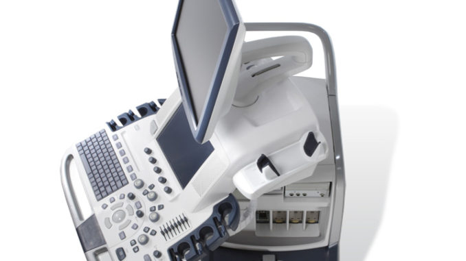
The Vivid* E9 is GE Healthcare’s first cardiovascular ultrasound system built specifically for 4D imaging — from ergonomics and image acquisition to data management and archiving. Vivid E9 provides the tools to help streamline your workflow, enhance productivity, and provide the data you need for confident diagnoses. Our Vivid E9 also provides exceptional 2D imaging with equal power, precision, and agility.
Raw Data Format
The GE Vivid product line has always acquired and stored its data in a specific raw data format, which enables onboard as well as after the fact post processing capabilities. This flexible and innovative format and storage of pre scan converted data has enabled development of utilities with high clinical value.
This has resulted in an enhanced ability to perform additional advanced algorithms for all of the steps in the data processing chain, culminating in the release of the Vivid E9 with XDclear*.
Extraordinary Image Quality
Crisp imaging. Focused workflow. Clear quantification. Vivid E9* makes short work of your routine 2D exams.
2D Image Quality
Designed with GE’s proprietary XDclear* technology, the M5Sc transducer delivers excellent endocardial definition and texture. Together with controls like UD Clarity and HD imaging, it helps provide crisp valves and borders across a wide range of patients.
Adult Echo Color Doppler: The XDclear transducer technology provides the color sensitivity needed for visualization of pulmonary vein inflow.4D Image Quality
The Vivid E9 helps make 4D imaging every bit as easy as 2D imaging. It can bring enhancements to your entire 4D workflow process, thanks to fast, consistent reproducibility
4D Strain Quantification: Supports a new export format for ease of doing offline research.
Depth render map: Copper/Blue for enhanced shadowing, render continuity, render stability and depth perception.
2 Click Crop for TTE: Efficient way of obtaining any 4D view in live and/or replay.
FlexiZoom: No need for flipping or cropping, but flexibility to make further adjustments (rotation, translation) if desired.
Extraordinary Quantification
Vivid E9 is all about making imaging simple, intuitive, and quantifiable to help make your work easy and efficient.
Automated Function Imaging (AFI)
This software tool assesses and quantifies left ventricular wall motion at rest. It calculates a large set of parameters to describe the function of the left ventricular walls. AFI specifically calculates peak systolic longitudinal strain (both segmental and global) and presents the results as parametric images. As part of its healthymagination validation, a study has shown that AFI offers potential in predicting mortality in patients with suspected LV impairment compared to Ejection Fraction and Wall Motion1.You can view the AFI healthymagination Fact Sheet and the AFI healthymagination Validation Summary.
1. Stanton et al, ‘Prediction of all-cause mortality from global longitudinal speckle strain: Comparison with ejection fraction and wall motion scoring’, Circulation: Cardiovascular Imaging, 2009; 2: 356-364
AFI: Bullseye as well as segmental traces showing reduced longitudinal strain, likely due to right coronary flow obstruction
4D Strain: As an extension to the 4D LV Mass tool, both global and regional strain values are calculated based upon a spatial speckle tracking algorithm. The end result is presented in a Strain Bull’s Eye plot accompanied by time-strain curves and cut planes for enhanced visual tracking assessment.
4D Auto LVQ: A mesh-based surface-tracking model, the 4D Auto LVQ quantification tool provides you with a graphical output of pure 4D volume data. Utilizing temporal data, it delivers reproducible results for automatic volumes and ejection fractions.4D LV Mass: Using the above-mentioned mesh-based surface tracking model, by adding the epicardial border, an LV Mass and an LV Mass Index is derived from the same data set.
MV Assessment: The semi-automated MV Assessment tool now also available on Vivid E9 for TEE and TTE, provides the ability to include quantitative results for the mitral valve apparatus, into the patient exam.
Extraordinary Workflow
The Vivid E9 helps make 4D imaging as easy as 2D imaging. It can bring remarkable enhancements to your entire 4D workflow process, thanks to fast, consistent reproducibility.
Scan Assist
With Scan Assist, you can quickly customize the system for your departmental protocols for CRT optimization, and let the system guide you to the next view, mode, and measurement.
4D Views: Used to automatically derive and visualize a conventional apical long axis view, in 4D as well as in two orthogonal slices.
SAX 4D stress views: Helping improve workflow for stress echo procedures, 4D Stress is an innovative first step in helping you integrate 4D into the routine of your day-to-day clinical practice.
FlexiSlice: Easily switched from volumes to slices and back in live or replay mode.
FlexiZoom: See the mitral valve in 4D TEE with one push of a button.
2-Click Crop: Click and drag. See any 4D view in live mode or replay.
Multi-slice: Available in live or replay, helps the user extract conventional long axis and short axis views from 4D volume datasets.
Vivid E9* with XDclear* delivers advanced technology and innovative transducers to image pediatric patients. XDclear technology helps you acquire images on a wide range of pediatric and neonatal patients, quickly and with minimal system adjustments. With XDclear transducers, you can image deep anatomy without sacrificing clarity.
Pediatric imaging
Two dedicated pediatric transducers, the 6S-D and 12S-D, cover patients ranging from neonates up to children weighing 16-18 kg. Vivid E9 with XDclear continues to provide the same exceptional 2D imaging power, precision and agility, along with color flow and Doppler. The new imaging presets for these transducers enhance low-level signal display in 2D and M-Mode. They combine high color sensitivity with high frame rates, by leveraging parallel beam forming power. The result is enhanced differentiation of small and fast moving structures and flows
Pediatric Imaging: The new setup of the 12S-D transducer combines high spatial and temporal resolution in both 2D and Color, as shown in this Simultaneous Mode image of a complex anatomy.
Neonatal and fetal heart imaging
With the introduction of Vivid E9 with XDclear, three new transducers are available for neonatal and fetal heart imaging. A new micro convex transducer, the 8C, is well-suited to head and abdominal imaging, with its tight lens curvature.
Neonatal head imagingFor fetal heart imaging, two new curved array transducers, the C1-5-D and C2-9-D, cover a range of fetuses, from early to late trimesters. The C1-5-D delivers excellent 2D and color flow imaging for late trimester fetal heart imaging. The new high frequency C2-9-D transducer employs the innovative XDclear technology and is well suited for early trimester imaging. Both transducers now use the full processing power of the beam former to help improve color flow visualization.
Fetal Heart imaging: Using the new XDclear C2-9-D high frequency curved array transducer for enhanced color flow sensitivity and resolution.
Depth Illumination
GE has standardized Depth Illumination on this platform to enhance communication to non-echo experts. The new depth color map helps visualization when the object is illuminated by an imaginary light source, casting a shadow enhancing depth/distance perception. This can be very helpful for an interventional device/catheter.
Example of ASD closure device visualized by help of Depth Illumination.
Polar Vision
Precise, efficient communication between the echocardiographer and the surgeon in the OR or the interventionalist in the cath lab is a prerequisite for a smooth procedure in a crowded and stressful environment. There is a continuing need for enhancements in 4D visualization of structures.
Vivid E9* with XDclear* helps provide detailed anatomical insight. It delivers extraordinarily clear depth perception through an innovative visualization technique called Polar Vision, that combines polarized stereo with depth rendering on a dedicated 3D monitor.
The Polar Vision option is designed to enhance communication in the cath lab and OR, introducing a new stereo vision technology combining polarized stereo with depth rendering displayed on a dedicated 3D monitor.
MV Assessment: And now, with the TomTec** MV Assessment tool on board the Vivid E9 in addition to the EchoPAC*, we provide comprehensive visual and quantitative assessment for structural heart repairs in the echo lab or interventional lab.
Overview
While Vivid E9* is currently used most frequently in the echo lab, the availability of the 4D TEE transducer enables the system to also be used in clinical settings including the cath lab, and the operating room.
- GE’s 4D TEE transducer allows clinicians to view precise images of the heart during assessment/diagnosis performed in the echo lab, and to support invasive surgical procedures in the operating room, as well as image-guided procedures in the cath lab
- With the 4D TEE transducer, Vivid E9 can now be used as a visual aid when performing procedures like mitral valve repair, transcatheter aortic valve implantation (TAVI), atrial septal defect (ASD) closures and patent foraman ovale (PFO) closures
The Vivid E9 also includes several features that are new to GE cardiovascular ultrasound, and which are designed to enhance 4D imaging and workflow.

- The industry’s first configurable 4D TEE transducer
- A new tilt and rotate function
- A live “2-Click Crop” tool
Workflow enhancements can also be achieved during mitral valve acquisition with a dedicated MV button and during quantification with a new plug-in tool that has the potential to help decrease assessment time.
Simplified Image Acquisition.
- Triplane imaging, 4D views, and high frame rates for both TTE and TEE procedures
- One-button acquisition of mitral valve images
- In biplane mode, the ability to tilt and rotate at the same time, e.g. to accurately image the mitral valve during a MV clip procedure
- A new QuickRotate button helps provide consistency and accuracy by allowing users to rotate an image in 30 degree increments with a single click
- Enhanced zoom capabilities allow for high volume rates in a single- and multibeat in 4D and 4D color zoom mode using FlexiZoom
Mitral Valve from surgeon’s view utilizing Flexizoom
BiPlane Tilt and Rotate
Intuitive Navigation
2 Click Crop, a quick and intuitive way of obtaining any 4D view in both live and replay modes.
Laser lines, a new tool to help physicians understand the relationship between 2D slices, and 4D views, help to visualize the linkage between 2D and 4D, and also helps improve depth perception
Innovative slice tool, FlexiSlice, provides flexibility for the user to slice in any direction, 2D view or render view in both live and replay modes.
Advanced Quantification
MV Assessment plug-in from TomTec** (Available on EchoPAC*)
- Helps increase productivity during mitral valve assessment
- Provides a set of static and dynamic measurements describing the mitral valve anatomy
An automated left ventricular volume and EF quantification tool (4D Auto LVQ) also for TEE
4D Auto LVQ for TEE


 The new Carotid_A preset provides enhanced 2D image quality in terms of spatial- and contrast resolution.
 The 8C probe with its Carotid application: has the tight curvature to overcome vascular access limitations on certain patients.
 The C2-9-D XDclear convex probe: combines high resolution and penetration as shown in this abdominal image.
 The C1-5-D convex probe: provides the sensitivity needed for renal flow as demonstrated in this case where Angio imaging is used.