
Visualization of the Red Reflex
The Stereo Coaxial Illumination (SCI) allows you to benefit from the detail recognition, high-contrast brilliance and stability of the red reflex – even with strongly pigmented, decentered and ametropic eyes. This technology enables you to see all the details of a patient’s eye.

Effortless positioning
The magnetic brakes make positioning the surgical microscope very simple. When the brakes are released, the system smoothly glides into a new position; when locked, the surgical microscope remains firmly in place.

Independent second view
The OPMI Lumera® T surgical microscope can be equipped with a completely integrated assistant’s microscope. The second surgeon selects the focus and magnification independently of the main surgeon, thus enabling active assistance.

Natural color impression
The integrated Superlux® Eye xenon illumination allows you to see the anatomic structure of the eye in its natural colors and highly accurate detail. The use of the HaMode™ filter allows surgeons who prefer halogen to quickly switch to a light spectrum equivalent to halogen. This is particularly beneficial when several surgeons with different preferences regarding the light source use one system.
External Video Components
To support all requirements or wishes for customized video solutions, external components can be mounted to the surgical microscope system. The external attachment via standard optical and mechanical interfaces increases the flexibility and retrofitability of the surgical microscopes.
Video Cameras

TRIO 610 with CCU TRIO 600 – 3 Chip HD Camera System
The high definition camera system with apochromatic video optics allows surgical microscope images to be generated with enhanced resolution and color rendition. The camera can be used for information, documentation, teaching and presentation of high quality images.

Ceiling mount
Spans large distances and features a smooth, lift function that makes it possible to quickly switch between the working and park position. This means you have a lot of headroom, both in the working position and the extremely compact park position.

RESIGHT 700 for retina surgery
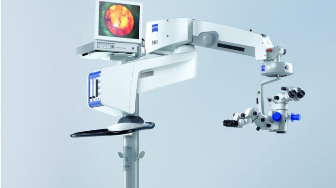
OPMI Lumera T and RESIGHT® 700 fundus viewing system allow you – the retinal surgeon – to clearly recognize every detail of the retina. And as these products work together seamlessly, they offer unparalleled convenience in the OR.

Stereo co-observation tube
OPMI Lumera T can be equipped with a stereo co-observation tube. This enables a second person to see the surgical field at the same magnification level. This is particularly well suited for sterile assistants or for training.

Foot control panel
It’s time to give you and your team more free space in the OR. Experience the new dimension of freedom with no annoying cables and free positioning in your OR. The ergonomically designed foot control panel (FCP and FCP WL) allows you to control your surgical microscope – intuitively and reliably. Additionally to our wireless version, we also offer a wired foot control panel.
| Surgical microscope | Apochromatic optics |
| Motorized zoom system, 1:6 zoom ratio | |
| Focusing range: 50 mm | |
| Binocular tube: Invertertube® (optional 0-180° tiltable tube) | |
| Objective lens f=200 mm (f=175 mm optional) | |
| DeepView: depth of field management system | |
| Integrated fully stereoscopic assistant’s microscope with no light loss for the main surgeon | |
| Illumination | Stereo Coaxial Illumination (SCI): red reflex illumination and full field illumination, patent pending |
| Light source | Superlux Eye xenon light source with manual bulb change, including HaMode filter |
| Halogen light source with fully automatic bulb change in case of a lamp failure | |
Option: Dual light source:
| |
| Fiber optic illumination | |
| Integrated 408 nm UV barrier filter | |
| Blue blocking filter | |
| Retinal protection device | |
| Optional: fluorescence filter | |
| X-Y coupling | 40 mm x 40 mm adjustment range |
| Reset-Button for X-Y coupling and focus | |
| Suspension systems |
|
| Maximum load: 20 kg (44.1 lb) (complete microscope equipment, including accessories) |
